How Nuclear Medicine Works: An Insight into Advanced Diagnostic Techniques

In the ever-evolving landscape of healthcare, diagnostic imaging has become an indispensable tool for identifying and managing a wide variety of medical conditions. One of the most advanced and specialized branches of imaging is nuclear medicine. It provides healthcare professionals with critical insights into how organs and tissues are functioning, rather than just revealing their structural details. Nuclear medicine is an essential tool in diagnosing a range of conditions, including cancer, heart disease, and neurological disorders.
At PRP Diagnostic Imaging, we are at the forefront of offering state-of-the-art nuclear medicine services. In this blog, we’ll delve into the question: how nuclear medicine works, exploring the technology behind it, its applications in healthcare, and how it helps improve patient outcomes.
Understanding Nuclear Medicine: The Basics
Nuclear medicine is a specialized area of medicine that uses small amounts of radioactive material (radiopharmaceuticals) to diagnose and treat various diseases. Unlike other imaging techniques that focus primarily on the structure of the body, nuclear medicine provides functional information about organs and tissues, allowing doctors to see how well a particular organ is working.
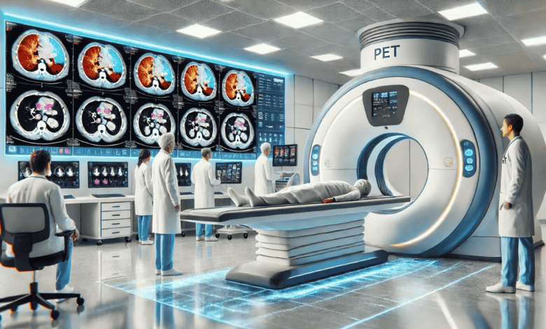
In nuclear medicine, a radiopharmaceutical is administered to the patient. This material is typically injected into a vein, swallowed, or inhaled. Once inside the body, it is absorbed by specific organs or tissues. The radiopharmaceutical emits radiation that can be detected by specialized cameras, such as a gamma camera, or by other imaging devices like PET (Positron Emission Tomography) and SPECT (Single Photon Emission Computed Tomography).
These imaging devices capture the emitted radiation and create images that help doctors assess the functioning of organs and tissues. The images produced by nuclear medicine scans reveal not just anatomical structures, but also the metabolic activity and function of these structures, which is crucial for diagnosing many types of diseases.
We utilize the latest nuclear medicine technology and equipment, ensuring that each patient receives the highest quality care and the most accurate diagnostic results.
How Nuclear Medicine Works: The Process
The process of nuclear medicine typically involves three main steps: the administration of the radiopharmaceutical, imaging, and interpretation of the results. Here’s a breakdown of each stage:
1. Administration of the Radiopharmaceutical
The first step in a nuclear medicine procedure is the administration of a small amount of radioactive material, or radiopharmaceutical, to the patient. The exact type of radiopharmaceutical used depends on the nature of the test and the organs or tissues being examined.
- Injected – Many nuclear medicine tests involve injecting the radiopharmaceutical into a vein. For example, in a bone scan, the radiopharmaceutical is injected into the bloodstream and is absorbed by bones.
- Swallowed – Some tests, like a thyroid scan, involve swallowing the radiopharmaceutical in pill or liquid form.
- Inhaled – In rare cases, the radiopharmaceutical is inhaled to assess lung function or other organs.
The radiopharmaceutical is designed to target specific organs, tissues, or cells, allowing for more precise imaging and diagnosis.
2. Imaging
Once the radiopharmaceutical has been absorbed by the body, the patient undergoes imaging. This is where the specialized equipment comes into play. Depending on the test, a gamma camera, PET scanner, or SPECT scanner is used to detect the radiation emitted by the radiopharmaceutical.
- Gamma Camera – The gamma camera is used in many nuclear medicine procedures to detect gamma radiation emitted from the radiopharmaceutical. It produces two-dimensional images of the distribution of the radiopharmaceutical in the body.
- PET and SPECT Scanners – Positron Emission Tomography (PET) and Single Photon Emission Computed Tomography (SPECT) scanners take these images to the next level by providing more detailed, three-dimensional views. These technologies are especially useful in assessing metabolic activity in organs and tissues, such as detecting cancer, heart disease, and brain abnormalities.
We employ the latest PET and SPECT technology, ensuring the highest-quality images and precise diagnostic results.
3. Interpretation of Results
Once the images are captured, the next step is the interpretation of the results. Nuclear medicine specialists, like those at PRP Diagnostic Imaging, are trained to read and analyze the complex images produced during the scan. The goal is to look for abnormal areas where the radiopharmaceutical has accumulated or been absorbed differently, which may indicate the presence of disease.
For example, in the case of cancer, the radiopharmaceutical may accumulate in areas where cancer cells are present, as these cells often have a higher metabolic rate. In heart disease, areas of the heart that are not receiving enough blood and oxygen will show up as “cold spots” on the scan.
Once the nuclear medicine specialist analyzes the images, they will provide a report to the referring doctor, who will use the results to determine a treatment plan.
Applications of Nuclear Medicine in Healthcare
Nuclear medicine is a versatile and powerful tool used to diagnose and treat various conditions. Below are some of the most common applications of nuclear medicine in modern healthcare:
1. Cancer Diagnosis and Staging
Nuclear medicine plays a pivotal role in oncology, particularly in cancer diagnosis, staging, and monitoring treatment efficacy. PET scans are commonly used to detect the presence of cancer and to assess the extent to which it has spread. PET scans can highlight areas of abnormal metabolic activity, helping doctors identify both primary tumors and metastases.
We offer advanced nuclear medicine services for cancer detection, including PET scans and bone scans, allowing for precise staging and effective treatment planning.
2. Cardiology: Assessing Heart Function
In cardiology, nuclear medicine is invaluable for assessing heart function and identifying conditions like coronary artery disease, heart attacks, and heart failure. Myocardial perfusion imaging (MPI), which uses a radiopharmaceutical to evaluate blood flow to the heart, is one of the most widely used techniques in nuclear cardiology.
At PRP Diagnostic Imaging, our nuclear medicine specialists use MPI scans to provide essential information about the heart’s blood flow and muscle function, helping doctors decide on the best course of treatment for heart conditions.
3. Neurological Imaging
Nuclear medicine also plays a significant role in diagnosing neurological conditions, such as Alzheimer’s disease, Parkinson’s disease, and epilepsy. Specialized scans, such as dopamine transporter scans (DaT scans), can provide critical information about brain activity and dysfunction. This helps doctors diagnose and monitor neurodegenerative conditions, enabling earlier intervention and better treatment outcomes.
4. Bone Scans for Metastasis and Infections
Bone scans are used to detect conditions such as bone infections, arthritis, and metastatic cancer. A radiopharmaceutical is injected into the bloodstream, and areas of the bone that are infected or affected by cancer show up as “hot spots” on the scan.
At PRP Diagnostic Imaging, we offer detailed bone scans to help doctors diagnose bone-related conditions, ensuring that appropriate treatments are started as soon as possible.
5. Infection and Inflammation Imaging
Nuclear medicine can also be used to detect infections and inflammation throughout the body. This is particularly useful in cases where the source of the infection is difficult to locate using traditional imaging techniques. Scans like the gallium scan can help identify areas of infection and inflammation, providing essential information for treatment.
Why Choose PRP Diagnostic Imaging for Nuclear Medicine?
We pride ourselves on providing cutting-edge nuclear medicine services that enable accurate diagnoses and informed treatment decisions. Here’s why you should choose us:
- Expert Nuclear Medicine Specialists: Our team of highly trained specialists is dedicated to providing precise imaging results, ensuring that your health is in the best hands.
- Advanced Technology: We use the latest PET and SPECT technology, ensuring that our diagnostic images are of the highest quality and accuracy.
- Comprehensive Services: Whether you need imaging for cancer, heart disease, or neurological conditions, we offer a full range of nuclear medicine services to address your needs.
- Patient-Centered Care: We prioritize patient comfort and care, ensuring a smooth and supportive experience throughout the imaging process.
Conclusion
Nuclear medicine is a vital and advanced diagnostic technique that provides healthcare professionals with critical insights into organ function and metabolic activity. By understanding how nuclear medicine works, patients can better appreciate the value of this specialized imaging and how it helps in the early detection and treatment of various conditions.
At PRP Diagnostic Imaging, we are committed to offering the most advanced nuclear medicine services, powered by expert specialists and the latest technology. Contact us today to learn more about how our nuclear medicine services can support your health and well-being.


